Applying Monitoring Electrodes
4-4 Monitoring the ECG
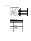
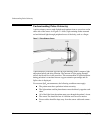
Figure 4-2 Precordial Lead Electrode Placement
Table 4-2 5-Lead ECG Lead Formation
Lead Lead Formation
ILA - RA
II LL - RA
III LL - LA
aVR
RA -
aVF
LL -
aVL
LA -
V
x
(or C
x
)
where x = 1-6
V/C -
1
2
3
4
5
6
LA + LL
2
RA + LA
2
RA + LL
2
RA + LA + LL
3
Electrode Location
V1 C1 forth intercostal space, at right sternal
margin
V2 C2 forth intercostal space, at left sternal
margin
V3 C3 midway between V2/C2 and V4/C4
V4 C4 fifth intercostal space, at left midclav-
icular line
V5 C5 same level as V4/C4, on anterior axil-
lary line
V6 C6 same level as V4/C4, at left mid axil-
lary line


